Best view resolution 1024x768
Online request for Sample Submission
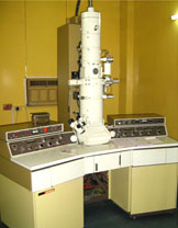
TRANSMISSION ELECTRON MICROSCOPE (TEM)Model: JEM-100 CX II
(Installed in 1983)

Applications:
Materials Science/Metallurgy, Biological Sciences, Medical Sciences, Nanotechnology, Ceramics, Pharmaceuticals, Semiconductors etc.
Specifications:
| Resolution: | 3Åto 1.4Å |
| Accelerating Voltage: | 20-100 KV in 20 KV steps. |
| Magnification: | With standard specimen gives 1000X to 450000X. In the low magnification mode it gives 100X, 200X, 400X and 600X. |
| Specimen Stage: | There is an airlock mechanism for the specimen exchange and it is provided for loading two specimens at a time. Specimen movement range is (±) 1 mm (built in specimen position indicator (CRT) for easy selection of desired field of view. |
| Instructions to users: | The specimen should be fixed in 2-3% glutaraldehyde in 0.1M Sodium cacodylate or phosphate buffer. Size of the biological tissue samples should be in the range of 1-2 mm. For material science samples, users may write for specific instruction according to sample type(s). |
| Accessories: | |
| Biological samples: | Ultra microtome(s), Knife maker |
Contact Tel. No. : 0364 2721806; 0364 272 1815.