Best view resolution 1024x768
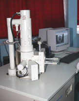
SCANNING ELECTRON MICROSCOPE (SEM)
Model: JSM-6360 (JEOL)
(Installed in 2004)
Model: JSM-6360 (JEOL)
(Installed in 2004)

Applications:
Entomology: Surface microstructure, systematics, development of sensory structure; Fish Biology: Surface microstructure of gills, scale, skin, gut etc. in relation to adaptation, environmental pollution, pathological conditions etc.; Parasitology: General morphology, surface microstructure etc. in relation systematics and drug efficacy.; Cell biology: Detail studies on cell-organelles with reference to physiology, metabolism, drug-delivary etc.; Limnology: Micro-structural studies on phytoplankton, zooplankton, eggs of different microarthopods etc.; Development biology: Surface micro-structural changes in relation to development.
Plant structures including leaf trichomes, wax bodies, pollen grains, seeds floral parts, fungal spores, hyphae etc.
Physical sciences: Characterization of nano particles, determination of crystal shape, angles and orientation, determination of grain size in rock materials etc.
Specifications:
| Resolution: | 3 nm in the secondary electron mode at working distance 8 mm and an accelerating voltage 0f 30 KV. |
| Accelerating Voltage: | From 1KV-30KV in 1KV step. |
| Magnification: | 8X (WD 48mm) to 300000X. |
| Image Modes: | SEI, BEI. |
| Maximum loadable Specimen size: | 10mm. |
| Accessories: | |
| Biological samples: | Critical point drier, Ion sputter system, carbon coating unit. |
| Material science samples: | Electromet 4 power source, Electromet 4 Polishing cell, Isomat low speed saw. |
| Instructions to users of SEM: | |
| Hard tissue: | Samples may be in fresh state. |
| Soft biological tissues: | Samples should be fixed in 2-3% glutaraldehyde in 0.1M Sodium cacodylate buffer. |
Contact Tel. No. : 0364 2721805; 0364 272 1818.